← Back to the plain-language case
Abstract reverses the paper's own stiffness ordering of intermediate EMT and mesenchymal cells
Commun Biol (2022) · doi:10.1038/s42003-022-03358-0 · PMC9061773
Summary
The abstract credits the intermediate (nest-like, partial-EMT) phenotype with "increased stiffness (and by inference invasiveness) ... compared to mesenchymal cells". The paper's atomic force microscopy (AFM) result says the opposite, and so do its Introduction, Results and Discussion: epithelial cells are the softest, mesenchymal cells the stiffest, and intermediate cells lie between them. The individual Young's modulus values in the paper's Supplementary Data 1 give means of 212, 560 and 980 Pa at 0 h (epithelial), 14 h (intermediate) and 48 h (mesenchymal), reproduce the three Fig. 5 P values, and put the intermediate-minus-mesenchymal difference at −420 Pa (95% CI −700 to −140). The measurements and the body text are consistent with each other; the abstract, whose only stiffness or invasiveness statement this is, reverses the direction of the measured effect and uses it as the basis for its translational statement.
What the paper reports
All quotations were checked verbatim against the PMC-archived text with src/show.py.
Abstract:
Further, owing to the increased stiffness (and by inference invasiveness) of the intermediate EMT phenotype compared to mesenchymal cells, our work can be instrumental in aiding the search for future treatment strategies that combat metastasis by specifically targeting the fiber alignment process.
Introduction, last paragraph:
Because the final aligned state is reported to have enhanced invasiveness, it would be advantageous to intervene at the intermediate states in order to prevent invasion.
We have also correlated the intermediate phenotype with lower stiffness compared to mesenchymal cells (stiffer than epithelial cells) supporting their partial EMT properties.
Results, section "The early EMT phenotype demonstrates a partial EMT nature and has different elastic properties compared to mesenchymal cells":
Due to the extensive interrelationship between the actin cytoskeleton and elastic properties of cells, the nest-like phenotype can also be expected to demonstrate a partial epithelial nature and be more compliant compared to the mesenchymal phenotype.
The epithelial cells had the lowest Young’s modulus and the late EMT mesenchymal phenotype had the highest Young’s modulus. The Young’s modulus of the nest-like phenotype was intermediate between the two (Fig. 5).
Fig. 5 legend:
Young’s Modulus values at 0 (blue) (n = 15), 14 (brown) (n = 13), and 48 (green) (n = 14) hours after EMT induction using TGFβ1 showing their mean and standard errors along with a plot of the mean values.
P1 = 0.0012, P2 = 0.0048, P3 = 3.6 × 10⁻⁹.
Methods, "Statistics and reproducibility":
We have carried out the two-sample t tests using the “ttest2” function in the Statistics and Machine Learning Toolbox in Matlab.
Discussion, first paragraph:
We demonstrated that the nest-like phenotype occupies an intermediate elastic niche between epithelial and mesenchymal cells.
As the cytoskeleton extensively affects the elasticity and motility of cells, we can expect the motility and invasiveness of the intermediate phenotype to be in between that of the epithelial and mesenchymal phenotypes.
Discussion, closing summary:
To summarize, in this work, we discovered a partial EMT phenotype in lung cancer cells with a unique cytoskeletal signature which are consistent with decreased stiffness compared to mesenchymal cells.
What the paper's own numbers show
Method. The main text gives no numeric moduli, so I used the paper's Supplementary Data 1 (42003_2022_3358_MOESM5_ESM.xlsx), whose sheet "Figure 5" lists each cell's Young's modulus in Pa at 0, 14 and 48 h after TGFβ1. For each time point I computed the mean, SD and SE. I then ran pooled-variance two-sample t tests (the default of Matlab's ttest2, named in the Methods) and pooled-variance 95% confidence intervals for the differences in means, and compared the results with the Fig. 5 legend and with the direction each passage states. recompute.py.txt holds the 42 values and prints every number below; given the .xlsx path, it also re-reads the file and checks the values against its copy.
| Quantity | Location | Reported | Recomputed from the paper's own numbers |
|---|---|---|---|
| Direction, intermediate vs mesenchymal | Abstract | "increased stiffness ... compared to mesenchymal cells" (EI > EM) | EI < EM: 560.0 vs 980.2 Pa |
| Direction, intermediate vs mesenchymal | Introduction; Results; Discussion (twice) | lower; more compliant; intermediate; decreased (EI < EM) | EI < EM (agrees) |
| Sample sizes | Fig. 5 legend | n = 15, 13, 14 | 15, 13, 14 values in Supplementary Data 1 |
| Young's modulus, 0 h (epithelial) | Fig. 5 (bars only) | not given in text | mean 212.4 ± 15.5 Pa (SE; SD 60.0) |
| Young's modulus, 14 h (intermediate, nest-like) | Fig. 5 (bars only) | not given in text | mean 560.0 ± 101.0 Pa (SE; SD 364.0) |
| Young's modulus, 48 h (mesenchymal) | Fig. 5 (bars only) | not given in text | mean 980.2 ± 91.5 Pa (SE; SD 342.4) |
| P1, 0 h vs 14 h | Fig. 5 legend | 0.0012 | 0.00115 (t = −3.65, df 26); EI − EE = +347.6 Pa, 95% CI 151.9 to 543.2 |
| P2, 14 h vs 48 h | Fig. 5 legend | 0.0048 | 0.00485 (t = −3.09, df 25); EI − EM = −420.2 Pa, 95% CI −700.2 to −140.2 |
| P3, 0 h vs 48 h | Fig. 5 legend | 3.6 × 10⁻⁹ | 3.6 × 10⁻⁹ (t = −8.56, df 27); EM − EE = +767.8 Pa, 95% CI 583.7 to 951.9 |
| Ratio EI / EM | — | abstract implies > 1 | 0.571 (the intermediate mean is 57% of the mesenchymal mean) |
| Intermediate cells below the mesenchymal mean (980.2 Pa) | — | — | 12 of 13 |
The source data reproduce all three legend P values to the printed precision, so they are the data behind Fig. 5. Unequal-variance (Welch) tests give P = 0.0049, 0.0050 and 1.05 × 10⁻⁶, and Mann-Whitney tests give P = 2.3 × 10⁻⁵, 0.0015 and 5.1 × 10⁻⁶. Under every test the intermediate phenotype is significantly less stiff than the mesenchymal phenotype, and the 95% CI of the difference lies entirely below zero. Of the five passages that state a direction, the abstract is the only one that contradicts these data.
Figure
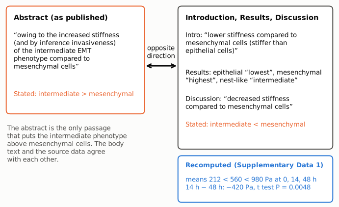
Figure 1. The conflicting statements. Left (orange frame): the abstract excerpt, which states that the intermediate phenotype is stiffer than mesenchymal cells. Right: short excerpts from the Introduction, Results and Discussion, which all place the intermediate phenotype below mesenchymal and above epithelial cells. The orange "Stated" lines give the direction each side reports. Bottom right (blue): values recomputed by recompute.py.txt from the paper's Supplementary Data 1. The mean moduli rise from 212 to 560 to 980 Pa, and the 14 h minus 48 h difference is −420 Pa with a pooled t test P value of 0.0048, the legend's P2. The source data agree with the body text and contradict the abstract.
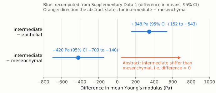
Figure 2. Differences in mean Young's modulus with pooled-variance 95% confidence intervals, recomputed from Supplementary Data 1 (blue), on one linear axis in Pa with zero as the reference line. Top row: intermediate minus epithelial, +348 Pa (95% CI +152 to +543), consistent with "stiffer than epithelial cells". Bottom row: intermediate minus mesenchymal, −420 Pa (95% CI −700 to −140). The orange arrow marks the side of zero the abstract's statement requires (difference > 0); the recomputed interval lies wholly on the other side.
Why it matters
- The abstract reverses a measured effect. This is the only stiffness or invasiveness statement in the abstract. It says the intermediate phenotype is stiffer than mesenchymal cells, but the AFM data show it is significantly softer (P = 0.0048), at 57% of the mesenchymal mean. A reader who sees only the abstract, or an indexing service or citing paper that relies on it, gets the opposite of the paper's finding.
- The reversed direction carries an inference about invasiveness. The abstract presents the intermediate phenotype as stiffer "and by inference" more invasive than mesenchymal cells. The paper's own reasoning in the Discussion runs the other way: invasiveness is expected "to be in between that of the epithelial and mesenchymal phenotypes", that is, lower than in mesenchymal cells. Corrected, the abstract's inference about invasiveness also changes direction. Invasiveness itself was not measured.
- The translational rationale rests on the reversed statement. The abstract gives the stiffness and invasiveness claim as the reason ("owing to") the work could help target fiber alignment to combat metastasis. The Introduction gives a rationale consistent with the data: the final aligned state "is reported to have enhanced invasiveness", so intervening at the intermediate states would be advantageous.
- The paper's main contributions are not affected. These are the SPOCC tool, the orientational order parameter as an EMT reporter, the split of EMT into stress-fiber formation followed by alignment, and the existence of a non-aligned intermediate phenotype. None of them depends on the abstract sentence. The AFM measurements, the statistics and the body text are internally consistent. The error is confined to the wording of the abstract.
Explanations considered
- A different population, time point or dataset. Does not account for it. There is a single AFM dataset: one TGFβ1 time course at 0 h (epithelial, n = 15), 14 h (nest-like or intermediate, n = 13) and 48 h (mesenchymal, n = 14). Supplementary Data 1 holds exactly these 42 values and reproduces the legend P values. No other comparison in the paper could make the intermediate phenotype stiffer than mesenchymal cells.
- "Intermediate EMT phenotype" means something else in the abstract. Does not account for it. The abstract uses the term for the intermediate EMT state it introduces in its second sentence, which the body calls the I or nest-like state. The abstract's words "compared to mesenchymal cells" rule out a comparison with epithelial cells, against which the intermediate phenotype is indeed stiffer.
- Stiffness read as compliance (an inverse quantity). Does not account for it. The abstract says "stiffness" outright and ties it to invasiveness. The Results use "compliant" only for the expectation that the nest-like phenotype is softer than mesenchymal cells, which again means EI < EM.
- The body text, not the abstract, is wrong. Ruled out by the source data. The individual moduli give EI < EM with a 95% CI of −700 to −140 Pa, and 12 of the 13 intermediate cells lie below the mesenchymal mean.
- A text-conversion or typesetting artifact. Ruled out. The discrepancy is in words ("increased" versus "lower" or "decreased"), not in a sign, exponent or table cell. The publisher's HTML, the PMC page and the Europe PMC full-text XML carry the same wording.
- Wording carried over from an earlier version. This bears on how the sentence arose, not on whether it is correct. The bioRxiv preprint (doi:10.1101/2021.06.17.448784, version 1, 17 June 2021) has the same sentence with "hence" in place of "by inference" and "intermediate phenotype" in place of "intermediate EMT phenotype". In the Peer Review File, a reviewer quotes the sentence and asks only that the invasiveness inference be reworded; the revised abstract changed "hence" to "by inference", and the comparison direction stayed as it was. In the same file, a reviewer's summary describes the intermediate phenotype as "associated with decreased invasiveness", which fits the Results, not the abstract.
- A correction already issued. None found (see Status).
Status
- Version of record. The abstract, Introduction, Results and Discussion passages quoted above are worded identically in the publisher's HTML (nature.com/articles/s42003-022-03358-0, read after its cookie redirect), the PMC page and the Europe PMC full-text XML. Every quotation was also checked verbatim against the PMC-archived text with
src/show.py. - Supplements. The PMC XML lists six supplementary items: the Peer Review File, Supplementary Information, a description of additional files, Supplementary Video 1, Supplementary Data 1 (XLSX) and the Reporting Summary. Supplementary Data 1 and the Peer Review File were downloaded from static-content.springer.com and read. Supplementary Data 1, sheet "Figure 5", resolves the question numerically, as shown above. Its SHA-256 (b991028077911ce5b8d6259b1c87ee5f1e92e5362830d8e5fdca7f94afc5bf7e) is recorded in
recompute.py.txtso the copy can be checked. - Corrections and comments. As of 2026-10-24 I found no correction, erratum, retraction or expression of concern.
src/known.pyreturns no Crossref update relations, no PMC notices and no Europe PMC notices. The Crossref record has no update-to or updated-by entries; its relations list only the bioRxiv preprint and a Faculty Opinions review. The publisher's page shows no correction notice. On PubPeer, an automated DOI query to the API returned no record (HTTP 404), and a web search for the title together with "PubPeer" found no thread. - Sources. PMC article PMC9061773; Europe PMC full-text XML; publisher page https://doi.org/10.1038/s42003-022-03358-0; Supplementary Data 1 (
42003_2022_3358_MOESM5_ESM.xlsx) and Peer Review File (42003_2022_3358_MOESM1_ESM.pdf); bioRxiv preprint 10.1101/2021.06.17.448784 (via api.biorxiv.org); Crossref; Europe PMC; PubPeer.
Suggested correction
In the abstract, replace
Further, owing to the increased stiffness (and by inference invasiveness) of the intermediate EMT phenotype compared to mesenchymal cells,
with wording that matches Fig. 5 and the body text, for example
Further, as the intermediate EMT phenotype has decreased stiffness compared to mesenchymal cells (and by inference lower invasiveness),
The rest of the sentence may then need rephrasing so that its rationale (targeting fiber alignment to combat metastasis) follows from the corrected direction; the Introduction already gives such a rationale, that the aligned mesenchymal state is reported to be more invasive and that intervening at the intermediate stage would therefore be advantageous. Stating the group means in the text would also help readers, for example mean ± SE Young's modulus of 212 ± 15, 560 ± 101 and 980 ± 92 Pa at 0, 14 and 48 h, as computed here from Supplementary Data 1.