← Back to the plain-language case
Abstract reports reduced PKN1 and PKN4, but the Results report increased PKN2 and no PKN4
Nat Commun (2018) · doi:10.1038/s41467-018-04411-5 · PMC5998138
Summary
For the isogenic cell-line comparison, the abstract states that PCQ "was used to identify reduced levels of serine/threonine protein kinases PKN1 and PKN4 in CFBE41o- cells compared to HBE41o- cells". The Results and Fig. 7 instead describe a PKN1/PKN2/PKN3 cluster in which PKN2 was "three-fold more abundant in CFBE41o- cells" and confirmed by western blot, the evidence for PKN1 is "conflicting" even about its presence, and PKN4 is never mentioned outside the abstract. The paper's own data files agree with the Results: PKN2-specific peptides have a CFBE41o-/HBE41o- ratio of 3.000, the only peptide on the PKN1 node is shared with PKN2 and PKN3 (ratio 0.500), and no file contains PKN4. Because the abstract is the indexed summary and this sentence is one of its two biological examples, it gets both the proteins and the direction of the one validated kinase change wrong.
What the paper reports
Abstract:
PCQ was used to identify reduced levels of serine/threonine protein kinases PKN1 and PKN4 in CFBE41o- cells compared to HBE41o- cells and to elucidate that shorter proteoforms of full-length caspase-4 and ephrin B receptor are differentially expressed.
Results, "Analysis of individual protein clusters", paragraph 2. This paragraph is the only passage in the Results about the PKN proteins:
Finally, a serine/threonine-protein kinase (PKN) cluster comprising PKN1 (Q16512), PKN2 (Q16513), and PKN3 (Q6P5Z2) was identified. PKN2 (PRK2) shares 59% sequence identity with PKN1 and was three-fold more abundant in CFBE41o- cells (Fig. 7b). Western blot confirmed that PKN2 was more abundant in CFBE41o- cells when normalized to the expression levels of Na+/K+-ATPase (Fig. 7c and 7d, Supplementary Fig. 12).
A difference in PKN2 protein quantity may contribute to the altered cell morphology between HBE41o- and CFBE41o– cells in cell culture because bronchial epithelial airway cells require PKN2 to establish apical junctions32.
PKN1 is part of two incomplete protein pairs with either PKN2 or PKN3, which provide conflicting evidence for a potential presence of PKN1. While the incomplete protein pair with PKN2 shows discrepant ratio values for the peptide nodes (∆ ≥ 2-fold), the incomplete protein pair with PKN3 shows similar ratio values (∆ < 2-fold).
Fig. 7 legend, panels b and d:
b The protein cluster of serine–threonine kinase-related protein kinases PKN1, PKN2, and PKN3 is displayed.
d Western blot analysis was used to verify the difference in PKN2 expression levels between HBE41o– and CFBE41o– cells.
Data in the western blot represent independent biological replicates, CFBE41o–: n = 2, HBE41o–: n = 2
Results, "Comparison of two isogenic cell lines with PCQ", on the labelling, which fixes the direction of the ratios:
CFBE41o– and HBE41o– cells were labeled with light and heavy isobaric isotopologues, respectively, and analyzed in a single experiment (n = 1)
Methods, "Cell culture and western blot". The only antibody against a PKN protein is anti-PKN2:
PKN2 and NaK-ATPase were detected by incubation with monoclonal anti-PKN2 antibody
Discussion. The summary of the cell-line comparison mentions no kinase:
When comparing two isogenic cell lines, we found that caspase-4 and ephrin type B receptor protein fragments were differentially regulated relative to their longer proteoforms, respectively.
What the paper's own numbers show
Method. I counted every mention of PKN1 to PKN4 in the PMC-archived text, separately for the abstract and for the rest of the article (Results, Discussion, Methods, figure legends). I searched every body sentence that names a PKN protein for wording of reduction ("reduc", "decreas", "lower", "less abundant", "down"). I then opened the two supplementary data files that hold the cell-line quantification. Supplementary Data 7 (MOESM9, the 180 classified complete protein pairs) gives log2 ratios for unique and shared peptide nodes. Supplementary Data 6 (MOESM8, "Network 5" in the Results) is the XGMML peptide-to-protein network for CFBE41o- vs HBE41o-, with ion counts for each peptide node. From it I extracted the connected component that holds the three PKN accessions. I converted log2 ratios to ratios. I checked the ratio direction against the normalized light and heavy ion counts, since the Results say light = CFBE41o- and heavy = HBE41o-. I also queried UniProt for a human gene named PKN4. Every number below is printed by recompute.py.txt.
| Quantity | Location | Reported | Recomputed from the paper's own text and data |
|---|---|---|---|
| Kinases named as changed in the cell-line comparison | Abstract | PKN1 and PKN4, "reduced levels" in CFBE41o- | Results name PKN2 as changed (increased); no reduction of any PKN protein is stated |
| Mentions of PKN1 / PKN2 / PKN3 / PKN4 | Abstract vs rest of article | 1 / 0 / 0 / 1 in the abstract | 5 / 13 / 4 / 0 in the rest of the article |
| Body sentences naming a PKN protein that use reduction wording | Results, Methods, legends | — | 0 of 11 |
| Members of the PKN cluster | Results; Fig. 7b legend | PKN1 (Q16512), PKN2 (Q16513), PKN3 (Q6P5Z2) | same three in Supplementary Data 6; no PKN4 node in the network file |
| PKN2-specific peptides (3 peptides), CFBE41o-/HBE41o- | Results ("three-fold more abundant in CFBE41o- cells"); Supp. Data 7 | log2 1.58496 | ratio 3.000 (= 2^1.585; normalized light/heavy ions 4.5/1.5 in Supp. Data 6), an increase |
| PKN2 western blot | Results; Fig. 7d (n = 2 vs n = 2); Supp. Fig. 12 | "more abundant in CFBE41o- cells" | an increase, the same direction as the mass-spectrometry ratio |
| PKN3-specific peptides (3 peptides) | Supp. Data 7 only | log2 −0.678 | ratio 0.625 (1.60-fold lower; light/heavy 2.5/4.0); not discussed in the text |
| Shared peptide IADFGLCK (PKN1, PKN2, PKN3) | Supp. Data 6, 7 | log2 −1.000 | ratio 0.500 (light/heavy 0.75/1.5) |
| Peptides mapped to the PKN1 node | Supp. Data 6 | — | only the shared IADFGLCK. The node groups Q16512 with two fragment entries (H0Y429, K7EL10), and its gene-name field reads "PKN1,PKN2" |
| PKN1–PKN2 incomplete pair | Results ("∆ ≥ 2-fold") | — | unique 3.000 vs shared 0.500, a 6.00-fold discrepancy |
| PKN1–PKN3 incomplete pair | Results ("∆ < 2-fold") | — | unique 0.625 vs shared 0.500, a 1.25-fold discrepancy |
| A PKN1-specific or PKN4 ratio | article and all supplementary files | — | none exists |
| Human gene named PKN4 | UniProt (query 2026-10-23) | — | none; human PKN genes are PKN1, PKN2 (alias PRK2, PRKCL2) and PKN3 |
The body's account is internally consistent, and it reproduces from the data files: both the "∆ ≥ 2-fold" and the "∆ < 2-fold" statements match the network ratios. The abstract sentence is the outlier. It names a protein (PKN4) with no data behind it in the paper. It describes as an identified reduction a PKN1 inference that the body calls conflicting. It leaves out the one PKN change that was quantified with protein-specific peptides and checked by western blot, which is an increase.
Figure
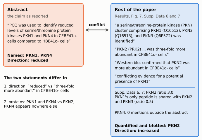
Figure 1. Short verbatim excerpts from the abstract (left, orange) and from the Results and Fig. 7 legend (right, blue), with the supplementary-data values printed by recompute.py.txt. The abstract names PKN1 and PKN4 and says "reduced". The body names a PKN1/PKN2/PKN3 cluster and reports PKN2 three-fold more abundant in CFBE41o- cells (ratio 3.0 in Supplementary Data 6 and 7), confirmed by western blot. It calls the evidence for PKN1's presence conflicting, and its only peptide is shared with PKN2 and PKN3 (ratio 0.5). PKN4 is mentioned nowhere outside the abstract.
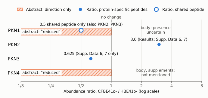
Figure 2. CFBE41o-/HBE41o- abundance ratios on one log2 axis; the vertical line marks no change (ratio 1). The orange hatched bands mark the abstract's claim for PKN1 and PKN4. The abstract gives a direction ("reduced") and no value, so the bands span the whole "reduced" side and carry no magnitude. Filled blue circles are ratios from protein-specific peptides in Supplementary Data 6 and 7: PKN2 at 3.0, which is the Results' "three-fold more abundant", and PKN3 at 0.625, which the text does not discuss. The hollow blue circle is the PKN1 node's only peptide, which is shared with PKN2 and PKN3 (0.5), so it is not specific to PKN1. PKN4 has no data point because the paper and its supplements contain none.
Why it matters
- The abstract misstates one of its two biological examples. The cell-line comparison is the paper's demonstration on a human proteome, and the abstract gives two results from it: the kinases and the caspase-4 and ephrin B receptor proteoforms. For the kinases, a reader of the abstract would conclude that PKN1 and PKN4 are lower in ∆F508-CFTR CFBE41o- cells. The body supports neither claim as stated. The one kinase result it quantified with protein-specific peptides and checked by western blot goes the other way: PKN2 is higher in CFBE41o- cells. The Results then offer an interpretation of that increase (apical junctions and cell morphology). The abstract's reduction cannot carry that interpretation.
- A protein name with no counterpart. PKN4 is not a human gene symbol in UniProt and has no entry in the article or its supplementary files. A reader who searches for PKN4 data, or indexes the abstract, finds nothing to match it.
- Certainty is overstated for PKN1. The abstract says PCQ "was used to identify reduced levels" of PKN1. The body says the two incomplete pairs "provide conflicting evidence for a potential presence of PKN1". The only peptide on the PKN1 node is shared with PKN2 and PKN3, and the PKN1–PKN3 pair's ratios (0.625 vs 0.500) are consistent with PKN3 alone. The mass-spectrometry comparison is a single labelled experiment (n = 1), and there is no PKN1 blot.
- What is not affected. The PCQ method, the two-species proof of principle (58% vs 21% completion of incomplete protein pairs), the caspase-4 and ephrin B receptor proteoform results, the PRDX analysis, and the body's PKN2 result do not depend on this sentence. The Discussion and the conclusions do not repeat it. The error is limited to the abstract, but it misstates which proteins changed and in which direction.
Explanations considered
- The ratios are defined the other way round (HBE41o-/CFBE41o-). This does not account for the discrepancy. The abstract fixes the comparison itself ("in CFBE41o- cells compared to HBE41o- cells"), and the Results use the same direction ("more abundant in CFBE41o- cells"). In the network file, light/heavy ion counts reproduce each stored ratio (4.5/1.5 = 3.000 for PKN2), and the Results state that CFBE41o- was the light label. So the ratios are CFBE41o-/HBE41o-, and PKN2 is increased in CFBE41o-. Even if the direction were flipped, the protein would be PKN2, not PKN1 and PKN4.
- "PKN4" is an alias of a protein in the body. This does not account for it. The aliases the paper and UniProt give for PKN2 are PRK2 and PRKCL2. UniProt lists no human gene named PKN4, and the cluster's three accessions are PKN1, PKN2 and PKN3.
- "PKN4" stands for PKN3, which is lower in the data. This would partly fit the numbers but is not supported by the text. PKN3's specific peptides have a ratio of 0.625 in Supplementary Data 7, a 1.6-fold decrease, which is below the paper's own 2-fold threshold. The Results describe PKN3 only as a member of the cluster, never as reduced. Even on this reading, the abstract would still leave out the validated PKN2 increase and overstate PKN1. The published material cannot show whether the abstract meant PKN3.
- The PKN1 reduction is a PCQ inference from the shared peptide. This is consistent with the data and could explain the PKN1 half of the sentence. The shared peptide IADFGLCK is at 0.500 while PKN2's specific peptides are at 3.000. Under the paper's logic, this 6-fold discrepancy suggests that another protein, possibly PKN1 (or the PKN2 fragment H0Y429 grouped in the same node), is present at lower levels. The body itself declines that conclusion, though, because the PKN3 pair "shows similar ratio values". The abstract therefore states as a finding what the body presents as unresolved. This does not account for PKN4, or for the omission of PKN2.
- The result is in the supplementary material. It is not. All supplementary files were opened, some in an earlier pass of this pipeline (see Status). Supplementary Information (MOESM2) holds the full western blot exposures (Supp. Fig. 12, PKN2 only) and does not otherwise discuss PKN. The PKN entries in Supplementary Data 6, 7 and 8 match the Results as tabulated above. The other network files either contain no PKN accession or belong to a different experiment, not the CFBE41o-/HBE41o- comparison. No file reports a PKN1-specific ratio, and no file mentions PKN4.
- A conversion or typesetting artifact. This is ruled out. The publisher's HTML carries the same abstract sentence, in the page body and in its metadata, as the PMC and Europe PMC versions. The discrepancy is in protein names and in the direction of change, not in symbols or digits.
Status
- Version of record. The publisher's page (nature.com/articles/s41467-018-04411-5, published 13 June 2018) has the abstract sentence word for word: PKN4 appears in the abstract text and in the page's metadata and nowhere else. The Results paragraph and the Fig. 7 legend match the PMC archive. Europe PMC's full-text XML carries the same text (prior check in this pipeline). Every quotation above was confirmed verbatim against the PMC-archived text with
src/show.py --grep. - Supplements. I downloaded Supplementary Data 7 (MOESM9, XLSX) and the network files (MOESM4–8, MOESM10–14, zipped XGMML) from the publisher's server and searched them for Q16512, Q16513, Q6P5Z2 and PKN4. The values used above come from Supplementary Data 7 and Supplementary Data 6. Supplementary Data 8 (Network 6) contains the same PKN component with identical ratios. An earlier pass in this pipeline opened the remaining files (MOESM1 PDF, MOESM2 Supplementary Information DOCX, MOESM3 XLSX) and found no PKN content apart from Supp. Fig. 12.
- Corrections and comments. As of 2026-10-23 I found no correction, erratum, retraction or expression of concern. Crossref lists no update relations for the DOI. PMC and Europe PMC list no correction or comment links (PMID 29899466). The publisher's page shows no correction notice, and a web search found none. I could not check PubPeer: its API and its search page both refused automated access (Cloudflare challenge; HTTP 403).
- Sources. PMC article PMC5998138; publisher page https://www.nature.com/articles/s41467-018-04411-5; supplementary files 41467_2018_4411_MOESM8_ESM.zip (Supplementary Data 6) and 41467_2018_4411_MOESM9_ESM.xlsx (Supplementary Data 7); UniProt REST (human PKN gene query); Crossref; Europe PMC.
Suggested correction
- Revise the abstract sentence so it matches the Results. For example: "PCQ was used to identify increased levels of the serine/threonine protein kinase PKN2 in CFBE41o- cells compared to HBE41o- cells, confirmed by western blot, and to elucidate that shorter proteoforms of full-length caspase-4 and ephrin B receptor are differentially expressed."
- If a statement about PKN1 is kept, word it as the body does: the evidence for a PKN1 proteoform is inconclusive and rests on a peptide it shares with PKN2 and PKN3.
- Remove "PKN4". If another protein was meant, name it and give the supporting data.