← Back to the plain-language case
Abstract swaps the retina and vitreous ranibizumab half-lives given in the Results and Discussion
Commun Biol (2020) · doi:10.1038/s42003-019-0731-9 · PMC6949284
Summary
The abstract reports half-lives for labeled ranibizumab of 3.34 days at the retina and 4.52 days in the vitreous. The Results and the Discussion both give the same two numbers the other way round: 4.52 days in the retina and 3.34 days in the vitreous. The fitted lines in Figure 6a give about 4.55 days for the retina and 3.29 days for the vitreous, so the paper's own figure agrees with the body text and the abstract has the compartment labels exchanged. The half-lives are the abstract's only quantitative result. With the abstract's labels, the vitreous value (4.52 days) falls outside the 2.6–4 day range of earlier non-human primate (NHP) studies that the Discussion uses to validate the method. With the body's labels (3.34 days) it falls inside.
What the paper reports
All quotations are verbatim from the article as archived in PMC. The superscript reference numbers that PMC flattens into the text ("days5,8,28", "ranibizumab29") are written here as superscripts.
Abstract:
Here, we show a proof-of-concept for labeled ranibizumab with observed half-lives of 3.34 and 4.52 days at the retina and in the vitreous, respectively.
Results, section "In vivo distribution of a long-acting anti-VEGF antibody in the eye", second paragraph:
Maximal ranibizumab concentrations were estimated to reach 0.1 and 0.028 mg/mL in the vitreous and retina, respectively (Fig. 6a). Further pharmacokinetic analysis showed that labeled ranibizumab exhibited a half-life and an area under the curve (AUC) of 4.52 days and 0.22 day × mg/mL in the retina. In the vitreous, the calculated half-life, AUC, clearance, and volume distribution were 3.34 days, 0.57 day × mg/mL, 0.44 mL/day, and 4.78 mL, respectively.
Discussion, second paragraph:
The pharmacokinetic analysis for ranibizumab showed a half-life of 3.34 days in the vitreous, which is consistent with half-life measured in NHPs (2.6–4 days5,8,28) and in humans for ranibizumab29 with invasive measures. By contrast, the half-life measured in the retina with this technology was higher than reported elsewhere (4.52 days as compared to 2.6–2.3 days from ref. 5).
Clearance and volume of distribution were also similar between both approaches (0.594 mL/day for clearance and 2.75 mL for volume of distribution from enzyme-linked immunosorbent assay (ELISA) as compared to 0.44 mL/day and 4.78 mL from ocular tracking).
Methods, "Data analyses":
The half-lives of labeled antibodies in the vitreous and at the retina were then measured using a non-compartmental analysis on mean concentrations for all data points tested in each compartment using at least five time points with time range between Day 2 and last Day tested as the curve fitting approach and an uniform weighting (WinNonlin, Pharsight Corp., Mountain View, CA).
From the vitreous results, clearance (CL) was calculated from the formula: CL = dose/AUC, where dose is the amount of protein injected IVT (0.25 mg)
The volume of distribution (V) was calculated from the equation: V = CL/kel, where kel is the elimination rate constant obtained from the slope of the terminal data from the concentration–time plot.
Figure 6 legend, panel a:
Pharmacokinetics in both vitreous and retina were then determined by non-compartmental analysis based on mean concentrations at 40% intensity.
What the paper's own numbers show
Method. I tabulated the half-life given to each compartment in the abstract, the Results and the Discussion. I then made three independent checks of which assignment is correct.
- Figure 6a plots log concentration against time for each compartment, with a fitted line. I measured the slope of each fitted line in the publisher's full-size image, calibrated against the panel's own axis ticks (decades on the y-axis, days 0 and 30 on the x-axis), and converted it to a half-life (t1/2 = ln 2 / kel).
- I compared each assignment with the literature ranges the Discussion cites.
- I checked the vitreous clearance and volume against the Methods formulas.
Every number below is printed by recompute.py.txt, which also re-measures the Figure 6a lines when given the image file.
| Quantity | Location | Reported | Recomputed from the paper's own numbers |
|---|---|---|---|
| Ranibizumab t1/2, retina | Abstract | 3.34 d | Results and Discussion give 4.52 d (mismatch) |
| Ranibizumab t1/2, vitreous | Abstract | 4.52 d | Results and Discussion give 3.34 d (mismatch) |
| Ranibizumab t1/2, retina | Results; Discussion | 4.52 d | the two sections agree |
| Ranibizumab t1/2, vitreous | Results; Discussion | 3.34 d | the two sections agree |
| Slope of fitted line, vitreous panel | Figure 6a | (not stated) | 0.0914 decades/day, kel 0.211/day, t1/2 3.29 d |
| Slope of fitted line, retina panel | Figure 6a | (not stated) | 0.0662 decades/day, kel 0.152/day, t1/2 4.55 d |
| Vitreous t1/2 vs NHP range 2.6–4 d | Discussion | "consistent with" | 3.34 d is inside the range; 4.52 d is outside it |
| Retina t1/2 vs ref. 5 (2.3–2.6 d) | Discussion | "higher than reported elsewhere" | 4.52 d is 1.74 times 2.6 d; 3.34 d would be 1.28 times |
| Vitreous clearance CL = dose/AUC | Results; Methods | 0.44 mL/day | 0.25 / 0.57 = 0.4386 mL/day (matches) |
| Ratio of the two half-lives | — | — | 4.52 / 3.34 = 1.353 |
Figure 6a resolves the conflict. The vitreous panel's fitted line falls by a factor of 10 about every 10.9 days, which gives a half-life of 3.29 days. The retina panel's line falls more slowly, by a factor of 10 about every 15.1 days, which gives 4.55 days. Both values are within 1.5% of the body-text numbers for the same compartment (1.42% and 0.64%). They differ from the abstract's numbers by 27% and 36%. The body text and the figure therefore agree, and only the abstract differs. Its two numbers are exactly the body's two numbers with the compartment labels exchanged.
Figure
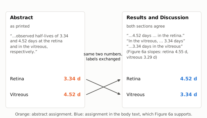
Figure 1. The two conflicting passages. Left: the abstract sentence and the assignment it states, retina 3.34 days and vitreous 4.52 days (orange). Right: the Results and Discussion, which both state retina 4.52 days and vitreous 3.34 days (blue). The arrows follow each number from one passage to the other: each one moves to the other compartment. The right panel also gives the half-lives implied by the slopes of the fitted lines in the paper's Figure 6a (4.55 days retina, 3.29 days vitreous), measured as described above. These agree with the body text.
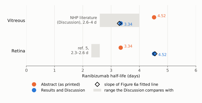
Figure 2. Ranibizumab half-life by compartment on a linear axis starting at zero. Orange dots: abstract. Blue dots: Results and Discussion. Open diamonds: half-life from the slope of the fitted line in Figure 6a. Grey bands: the literature ranges the Discussion compares each compartment with, 2.6–4 days in NHPs for the vitreous and 2.3–2.6 days from ref. 5 for the retina. The Figure 6a slopes sit on the body-text values. Under the body-text assignment, the vitreous value lies inside its literature band, as the Discussion says ("consistent with"). Under the abstract's assignment it lies outside.
Why it matters
- The abstract's only quantitative result is misattributed. The two half-lives are the only numbers in the abstract, and they are presented as the proof of concept for the method. A reader of the abstract, or of an index or database that copies it, takes away the opposite pairing to the one the paper's data support.
- It reverses which compartment clears more slowly. According to the body text and Figure 6a, the retina has the longer apparent half-life, 35% longer than the vitreous (4.52 / 3.34 = 1.35). The abstract implies that the vitreous does.
- It undercuts the validation argument as summarised in the abstract. The Discussion supports the imaging method by noting that the vitreous half-life is "consistent with" invasive NHP measurements of 2.6–4 days. This holds for 3.34 days and not for the abstract's 4.52 days. The Discussion's second point, that the retinal value exceeds tissue-extraction values, also weakens with the abstract's labels: 1.28 times the upper end of ref. 5 instead of 1.74 times.
- What is not affected. The values themselves, the body text, Figure 6a, the vitreous clearance (0.44 mL/day, which reproduces as 0.25 / 0.57), the free-dye half-lives (2.68 and 2.66 days), the Day 21 comparison of ranibizumab with the long-acting antibody, and the feasibility conclusion all stand. The error is confined to the labels in one sentence of the abstract.
Explanations considered
- A different analyte. Does not account for it. All three passages concern labeled ranibizumab. The free dye has its own half-lives (2.68 days vitreous, 2.66 days retina), and neither is 3.34 or 4.52.
- A different analysis, time window or definition of half-life. Does not account for it. The Methods and the Figure 6 legend describe a single non-compartmental analysis on mean concentrations (Day 2 to the last day, uniform weighting), applied to both compartments. The paper describes no second analysis that could produce the same two numbers with the compartments reversed.
- An ambiguous "respectively". Does not account for it. The abstract lists the values and the compartments in explicit order: "3.34 and 4.52 days at the retina and in the vitreous, respectively". It has only one reading.
- The body, not the abstract, is in error. Not supported. The Results and the Discussion agree with each other. The Discussion's reasoning only works with the body's assignment: it calls the vitreous value "consistent with" 2.6–4 days and the retinal value "higher than" 2.3–2.6 days. Most directly, the slopes of the fitted lines in Figure 6a give 3.29 days for the vitreous and 4.55 days for the retina.
- A text-conversion artifact. Ruled out. The publisher's HTML on nature.com has the same wording and numbers in the abstract, Results and Discussion, and so do the PMC text and the PubMed abstract.
- A correction already issued. None found (see Status).
- Side observation, not part of the finding. The reported vitreous volume of distribution, 4.78 mL, does not follow from the Methods formula V = CL/kel with either half-life. With CL = 0.44 mL/day it gives 2.12 mL for 3.34 days and 2.87 mL for 4.52 days. Reaching 4.78 mL would need a half-life of about 7.5 days (7.53 days). By comparison, the ELISA values from ref. 5 that the Discussion quotes (CL 0.594 mL/day, V 2.75 mL) imply 3.21 days, within the NHP range. The software may have used a different AUC internally (for example AUC extrapolated to infinity), so I do not treat this as an error, and it does not help decide which half-life belongs to which compartment. Separately, the "threefold vitreous:retina ratio" in the Discussion corresponds to an AUC ratio of 2.59 (0.57 / 0.22) and a peak-concentration ratio of 3.57 (0.1 / 0.028). This is within the looseness of the word "threefold".
Status
- Version of record. I downloaded the publisher's article page (https://www.nature.com/articles/s42003-019-0731-9). Its abstract contains "half-lives of 3.34 and 4.52 days at the retina and in the vitreous, respectively", and its Results and Discussion contain the passages quoted above, unchanged. The PMC HTML, the Europe PMC full text and the PubMed abstract (PMID 31925329) carry the same text. I checked every quotation verbatim against the PMC-archived text with
src/show.py. Figure 6a was measured in the publisher's full-size image of Figure 6. - Supplements. The publisher lists two supplementary files. The Supplementary Information (
42003_2019_731_MOESM1_ESM.pdf, 5 pages; Supplementary Figs. 1–4: dye comparison, free-dye localization, injection sites, calibration curves) contains no half-life, AUC or other pharmacokinetic values. The Reporting Summary (MOESM2) is a checklist. There are no source data. The Data availability statement says the datasets are available from the corresponding author on reasonable request. The supplements cannot settle the discrepancy, but the main-text Figure 6a does. - Corrections and comments. As of 2026-10-24 I found no correction, erratum, retraction or expression of concern.
src/known.pyreturns no Crossref update relations, PMC notices or Europe PMC notices. The Crossref record has no update-to or updated-by entries, the PubMed record has no CommentsCorrections entries, the Europe PMC record lists no comments or corrections, and the publisher's page shows no correction notice. The PubPeer API returned no record (HTTP 404) for the DOI, and a web search for the title together with "PubPeer" found no thread. - Sources. PMC article PMC6949284; https://www.nature.com/articles/s42003-019-0731-9 and its Figure 6 image and supplementary files; Europe PMC; PubMed (PMID 31925329); Crossref; PubPeer; https://doi.org/10.1038/s42003-019-0731-9.
Suggested correction
In the abstract, replace
observed half-lives of 3.34 and 4.52 days at the retina and in the vitreous, respectively
with
observed half-lives of 4.52 and 3.34 days at the retina and in the vitreous, respectively
which matches the Results, the Discussion and the fitted lines in Figure 6a. The authors may also wish to state how the vitreous volume of distribution of 4.78 mL was obtained. The Methods formula V = CL/kel gives about 2.1 mL with the reported clearance (0.44 mL/day) and vitreous half-life (3.34 days).