← Back to the plain-language case
Abstract reverses the direction of the paper's exposed-versus-occluded skin connectivity result
PLoS Comput Biol (2018) · doi:10.1371/journal.pcbi.1006099 · PMC5927471
Summary
The abstract says that skin sites exposed to the external environment have less connected phage-bacteria networks and are "likely more susceptible to network degradation". The Results say the opposite: occluded sites were less connected than exposed and intermittently occluded sites (p < 0.001, Fig 4B). The Discussion also says exposed and intermittently exposed sites were more connected and predicted to be more resilient. Fig 4B as published agrees with the Results and Discussion: the median eigen centrality is about 0.176 for exposed sites and 0.138 for occluded sites, and the paper reads higher values as "more connected" throughout. The body of the paper is internally consistent, so the analysis is unaffected. But the abstract, which is what most readers and indexing services see, states one of the paper's three headline findings in the reverse direction, and so does its claim about vulnerability to degradation.
What the paper reports
Abstract:
Network structure differed between skin sites, with those exposed to the external environment being less connected and likely more susceptible to network degradation by microbial extinction events.
Results, "Network structures across the human skin landscape", second paragraph:
The average centrality of each sample was quantified using the weighted eigenvector centrality metric. Intermittently moist skin sites (dynamic sites that fluctuate between being moist and dry) were significantly more connected than the moist and sebaceous environments (p-value < 0.001, Fig 4A). Also, skin sites that were occluded from the environment were less connected than those that were constantly exposed to the environment or only intermittently occluded (p-value < 0.001, Fig 4B).
Discussion, third paragraph:
Network structure differed between environmentally exposed and occluded skin sites. The sites under greater environmental fluctuation and exposure (the exposed and intermittently exposed sites) were more connected and therefore were predicted to have a higher resilience against network degradation when random nodes are removed from the network.
Together these data suggested that body sites under greater degrees of fluctuation harbored more highly connected microbiomes that are potentially more robust to network disruption by extinction events.
Fig 4 legend:
(A) Notched box-plot depicting differences in average eigenvector centrality between moist, intermittently moist, and sebaceous skin sites and (B) occluded, intermittently occluded, and exposed sites.
Sample sizes for each group were: Moist = 81, Sebaceous = 56, IntMoist = 56, Occluded = 106, Exposed = 61, IntOccluded = 26.
What the paper's own numbers show
Method. The paper prints no group values for connectivity in text or tables. They appear only as box plots in Fig 4A and 4B. I took the Fig 4 raster (3800 × 3729 px) embedded on page 11 of the publisher's PDF. For each box I located the median line and the two hinges (quartiles) by pixel row, and converted rows to eigen-centrality units with a straight line fitted to the panel's own y-axis ticks. The fit is exact to within 1e-4, and one pixel is about 0.0003 units, so each value is good to about ±0.002. I then checked the group sizes in the legend. I also took the pairwise Wilcoxon p-values that the authors' public code repository writes for this figure (rtables/skinboxsig.tsv; R's pairwise.wilcox.test, Holm adjustment by default). Finally I compared the direction each passage states with the direction the figure shows. The paper's convention, which the Results apply to both panels, is that a higher eigen centrality means "more connected". Every number below is printed by recompute.py.txt.
| Quantity | Location | Reported | Recomputed from the paper's figure and data |
|---|---|---|---|
| Exposed vs occluded connectivity | Abstract | Exposed sites "less connected" and "more susceptible to network degradation" | Exposed median 0.176 > occluded median 0.138: exposed sites are the more connected |
| Occluded vs exposed and intermittently occluded | Results; Fig 4B | Occluded "less connected" (p < 0.001) | Occluded 0.138 < exposed 0.176 and IntOccluded 0.185, which agrees |
| Exposed and intermittently exposed | Discussion | "more connected", "higher resilience" | Agrees with Fig 4B |
| Fig 4B medians (Q1–Q3) | Fig 4B | Box plots only | Exposed 0.176 (0.147–0.196); IntOccluded 0.185 (0.172–0.194); Occluded 0.138 (0.094–0.173) |
| Median differences | Fig 4B | Not printed | Exposed − Occluded = +0.038; IntOccluded − Occluded = +0.047; IntOccluded − Exposed = +0.009 |
| Separation of the groups | Fig 4B | Not printed | The exposed median (0.176) lies above the occluded upper quartile (0.173) |
| Fig 4A medians (Q1–Q3), the convention check | Fig 4A; Results | IntMoist "more connected" than Moist and Sebaceous | IntMoist 0.187 (0.175–0.197) > Sebaceous 0.145 (0.122–0.167) and Moist 0.137 (0.078–0.176), so higher means "more connected" |
| Group sizes | Fig 4 legend | 81 + 56 + 56; 106 + 61 + 26 | Both groupings sum to 193 samples, which is consistent |
| Occluded vs exposed p | Results; repository table | p < 0.001 | Holm-adjusted Wilcoxon p = 1.38e-05, which agrees |
| Occluded vs IntOccluded p | Results; repository table | p < 0.001 | p = 1.38e-05, which agrees |
| IntOccluded vs exposed p | Repository table | Not stated in text | p = 0.277 (no difference) |
The Results, the Discussion, the figure and the repository's p-values agree with each other. The abstract is the only place in the paper where exposed sites are described as less connected.
Figure
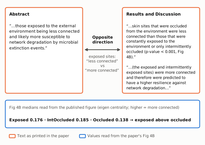
Figure 1. The left panel gives the abstract's sentence on exposed skin sites. The right panel gives the Results sentence on Fig 4B and the Discussion sentence on the same comparison. All three are quoted as printed and framed in orange. The connector marks the conflict: the abstract says exposed sites are "less connected", while the Results and the Discussion say they are more connected than occluded sites. The blue strip gives the Fig 4B medians that recompute.py.txt reads from the published figure. In those medians, exposed (0.176) and intermittently occluded (0.185) sites are above occluded (0.138) sites, which agrees with the body of the paper.
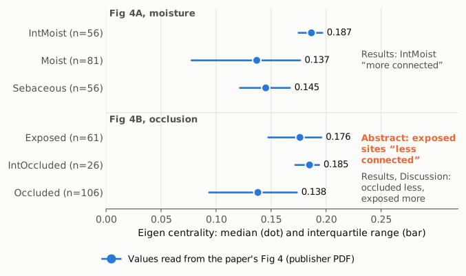
Figure 2. Median eigen centrality (dot) and interquartile range (bar) for the six skin-site groups. All values are read by recompute.py.txt from the published Fig 4A and 4B and are shown on one axis that starts at zero. The moisture panel (top) fixes the paper's convention: the Results call IntMoist, the highest group, "more connected". The occlusion panel (bottom) shows exposed sites above occluded sites. The orange label gives the abstract's statement, which runs the other way. Group sizes are from the Fig 4 legend.
Why it matters
- It is a headline finding. The abstract gives three findings from this study: networks are person-specific, high-fat diets go with less connected networks, and skin networks depend on environmental exposure. The third is printed with the wrong sign.
- The ecological inference is reversed too. The abstract says exposed sites are "likely more susceptible to network degradation by microbial extinction events". The Discussion draws the opposite conclusion from the same result: exposed and intermittently exposed sites are "predicted to have a higher resilience against network degradation". It builds its broader thesis on that conclusion, that "body sites under greater degrees of fluctuation harbored more highly connected microbiomes". A reader of the abstract alone would take away the opposite of the paper's conclusion.
- Abstracts travel further than full texts. Indexing services, citing papers and reviews usually quote the abstract. A study citing this paper for "exposed skin sites have fragile phage-bacteria networks" would be citing the reverse of what the data show.
- What is not affected. The analysis, Fig 4, the p-values, the Results and the Discussion are consistent with one another. The abstract's general framing still holds: network structure differs between skin sites, and environment may shape phage-bacteria interactions. Only the direction of the skin example is wrong.
Explanations considered
- The abstract compares exposed sites with a different group. It cannot. The occlusion grouping has only three levels. Exposed sites are above occluded sites (0.176 vs 0.138, p = 1.38e-05) and do not differ from intermittently occluded sites (p = 0.277). No comparison in the paper makes exposed sites less connected. The moisture grouping is a separate classification, and the abstract's sentence is explicitly about exposure to the external environment.
- Lower centrality means more connected. The paper never reads its values that way. The Results call the highest group in Fig 4A (IntMoist) "more connected" and the lowest group in Fig 4B (Occluded) "less connected". In the gut analysis, "reduced degree centrality" and "decreased closeness centrality" go with "less connected" networks. The authors' script (
bin/CompareSkin.R) computes the plotted value as igraph'scentr_eigen(...)$centralization, a graph-level eigenvector centralization score. I did not re-run that analysis. However that metric is best interpreted, the paper reads it one way in the Results and the Discussion, and the abstract states the opposite under that same reading. - The abstract summarises the supplementary dissimilarity analysis (Fig 4D and 4F). That analysis says only that "Occluded sites were significantly different from exposed and intermittently occluded sites". It compares the spread of network dissimilarities (ANOVA and Tukey test on group dispersion) and has no direction of connectedness. It cannot support "less connected".
- A conversion or typesetting artifact. Ruled out. The same abstract wording appears in the publisher's HTML and PDF, in the PMC copy and in the Europe PMC record.
- An error in reading the figure. It cannot change the conclusion. The direction comes from the paper's own Results and Discussion text. The figure reading only corroborates it. The exposed and occluded medians sit about 120 pixel rows apart on the 3800-pixel raster, a gap of 0.038 units against a reading precision of about 0.002.
- A later correction fixed it. None was found (see Status). Crossref has one update, a publisher "new version" entry dated 2018-04-30. It matches the switch from the uncorrected proof to the final version and does not change the abstract.
Status
As of 2026-10-23:
- Version of record. The abstract wording quoted above is in the publisher's HTML and PDF (24 pages, dated April 18, 2018), the PMC copy (PMC5927471) and the Europe PMC record (PMID 29668682). For this write-up, Fig 4 was read from the publisher PDF as held in the PMC open-data collection (
pmc-oa-opendata/PMC5927471.1/PMC5927471.1.pdf). - Supplements. S1–S14 Fig and S1–S2 Table are listed on the publisher page. S14 Fig only tests whether the Fig 4A and 4B p-values survive added edge noise, and gives no group values. The authors' public code repository, linked from the article, contains the analysis script (
bin/CompareSkin.R) and the p-value table used above (rtables/skinboxsig.tsv). It has no per-sample centrality values. - Corrections. No correction, erratum, expression of concern or retraction was found. Crossref has only the 2018-04-30 "new version" entry and a link to the bioRxiv preprint (10.1101/144642). PMC and Europe PMC list no notices. Europe PMC's comment/correction list holds only the preprint link.
- PubPeer. The PubPeer search API and site returned a human-verification challenge to automated requests, so a direct search could not be done. A web search for the article title on PubPeer found no PubPeer thread.
Suggested correction
In the Abstract, replace
Network structure differed between skin sites, with those exposed to the external environment being less connected and likely more susceptible to network degradation by microbial extinction events.
with
Network structure differed between skin sites, with those occluded from the external environment being less connected and likely more susceptible to network degradation by microbial extinction events.
or, stated from the exposed side,
Network structure differed between skin sites, with those exposed to the external environment being more connected and likely more resilient to network degradation by microbial extinction events.
Either wording matches the Results, Fig 4B and the Discussion. No other part of the paper needs to change.