← Back to the plain-language case
Abstract reports lighter liver copper isotopes in infected mice; the paper's own data show heavier
Commun Biol (2026) · doi:10.1038/s42003-025-09464-z · PMC12881638
Summary
The abstract says that Plasmodium berghei (Pb) infection gave a "significantly lighter Cu isotopic composition in the liver". The Results, the Discussion and the supplementary data all report the opposite: infected livers have higher δ65Cu than uninfected livers, and by the paper's own Eq. 3 a higher δ65Cu means isotopically heavier Cu. The per-mouse values in the Supplementary Data file give mean wet-liver δ65Cu of +0.09‰ in uninfected mice, +1.03‰ at 7 days post infection (DPI) and +0.65‰ at 10 DPI. Every one of the 16 untreated infected mice (7 and 10 DPI) lies above every control (Mann–Whitney p = 0.0007 at each time point). The abstract is where most readers meet this result, and the wrong sign there also makes the abstract's own mechanism (export of isotopically light Cu from the liver through ceruloplasmin) read as self-contradictory. The measurements, the statistics and the conclusions in the body of the paper are not affected.
What the paper reports
Abstract, fourth sentence:
A discrete hepatic Cu deficiency, but significantly lighter Cu isotopic composition in the liver were observed, alongside a release of Cu into the bloodstream through ceruloplasmin, indicating a contribution of Cu in potentiating Fe release from hepatic stores.
Results, section "Mice respond to infection with changes in the metal isotopic composition…", fourth paragraph (Fig. 3F, G):
As for Fe, also the isotopic compositions of Zn and Cu differ significantly between Pb-infected and uninfected mice (Fig. 3F, G). In wet livers, Pb infection led to lower δ66Zn values in comparison to uninfected mice, while it led to higher δ65Cu values compared to controls. Changes in the isotopic composition of Zn and Cu were also observed in perfused livers, albeit without statistical significance for Zn.
Results, same section, fifth paragraph:
Different to the observation for Fe, the largest absolute shifts in the isotope ratios of Zn and Cu were found at 7 DPI (onset of infection) rather than at 10 DPI (later stage).
Results, section "Host metal isotopic compositions are partly restored by antimalarial treatment", second and fourth paragraphs:
Similarly to Fe, the Zn and Cu isotope ratios shifted towards the values of uninfected mice as a result of antimalarial treatment (Fig. 4C, Table S3, Fig. S6).
Systematically, we found a negative correlation between δ56Fe values in the liver and the plasmatic concentration of hepcidin and a positive correlation between δ65Cu values in the liver and the plasmatic concentration of CP (Fig. 4D and E).
Discussion, eighth paragraph:
In parallel, a hepatic Cu deficiency was observed. Consistent to this deficiency, an increase in the heavy Cu isotope was noticed in Pb infection, where the highest increase has coincided with the onset of infection and anemia. We interpreted that when hepatocytes are gradually delivering Cu that is enriched in the light Cu isotope via CP into the plasma, then an enrichment in the heavier Cu isotope in the liver can occur. Antimalarial treatment has decreased the plasmatic levels of CP and the hepatic δ65Cu values, which is consistent with a profile of restoration of the Cu homeostasis.
Discussion, tenth paragraph:
In support to this, we have seen a hepatic Cu deficiency and a depletion in the lighter isotope, which is encompassed by an increased plasmatic Cu level.
Materials and methods, "Instrumentation and measurements…" (Eq. 3 is printed as δ65Cu = [(65Cu/63Cu)sample / (65Cu/63Cu)NIST SRM 976 − 1] × 1000):
δ value expresses the relative difference in the ratio of a heavy to a light isotope between a sample and an isotopic reference material. Therefore, a positive δ value indicates that isotopic fractionation has led to an enrichment in the heavier isotope in the sample compared to the reference material, while a negative δ value indicates a depletion in the heavier isotope.
Supplementary Information, Table S4 ("Metal isotope ratios in wet liver samples…", δ65Cu column, mean ± SD, ‰):
Untreated, 0 DPI 0.08 ± 0.12 · Untreated, 7 DPI 1.02 ± 0.29 · AQ 24 h, 10 DPI 0.37 ± 0.11 · CQ 24 h, 10 DPI 0.37 ± 0.11 · CQ 48 h, 10 DPI 0.31 ± 0.07 · Untreated, 10 DPI 0.65 ± 0.19
All quotations from the article were checked verbatim against the PMC-archived text with src/show.py; the Table S4 excerpt is from the publisher's Supplementary Information PDF (MOESM2), re-laid on one line.
What the paper's own numbers show
Method. First, the sign: from Eq. 3, the difference between two δ65Cu values is 1000 × (Rinfected − Rcontrol) / RNIST 976, where R is the 65Cu/63Cu ratio. Because the reference ratio is positive, a higher δ65Cu always means more 65Cu relative to 63Cu, that is, isotopically heavier Cu, whether the δ values themselves are positive or negative. Second, the size: I took the per-mouse liver δ65Cu values from the paper's Supplementary Data file (MOESM4, sheet "Liver"), recomputed each group's mean and SD, checked them against SI Tables S4 and S5, and compared infected with uninfected livers with the test the paper uses (two-sided Mann–Whitney, exact). The sheet labels the untreated infected groups "LP" and "HP" without a time point; their means and SDs match Table S4's 7 DPI and 10 DPI rows respectively, and that is how they are assigned here. Every number below is printed by recompute.py.txt.
| Quantity | Location | Reported | Recomputed from the paper's own numbers |
|---|---|---|---|
| Direction of the infection effect on liver Cu isotopes | Abstract | "significantly lighter Cu isotopic composition in the liver" | heavier: every body-text statement and every data source below gives higher δ65Cu in infected liver |
| Direction, same comparison | Results (Fig. 3G); Discussion paragraphs 8 and 10 | "higher δ65Cu values compared to controls"; "an increase in the heavy Cu isotope"; "a depletion in the lighter isotope" | consistent with each other and with Eq. 3: heavier |
| Wet liver δ65Cu, uninfected (0 DPI) | Table S4; MOESM4 | 0.08 ± 0.12‰ | +0.086 ± 0.124‰ (n = 6) |
| Wet liver δ65Cu, infected 7 DPI | Table S4; MOESM4 | 1.02 ± 0.29‰ | +1.025 ± 0.297‰ (n = 8); shift +0.94‰; 8 of 8 mice above every control; U = 48, p = 0.00067 |
| Wet liver δ65Cu, infected 10 DPI | Table S4; MOESM4 | 0.65 ± 0.19‰ | +0.650 ± 0.194‰ (n = 8); shift +0.56‰; 8 of 8 mice above every control; U = 48, p = 0.00067 |
| Perfused liver δ65Cu, 0 vs 10 DPI | Table S5; MOESM4 | 0.29 ± 0.25 vs 0.59 ± 0.17‰, p = 0.0306 | +0.309 ± 0.287 (n = 5; does not reproduce the printed 0.29 ± 0.25) vs +0.660 ± 0.263‰ (n = 9), shift +0.35‰, p = 0.042; without one infected row (see below) +0.591 ± 0.174‰ (n = 8), shift +0.28‰ |
| Treated (10 DPI) vs untreated 10 DPI | Results; Discussion p8; Table S4 | treatment "decreased … the hepatic δ65Cu values", toward uninfected | CQ 24 h +0.377, CQ 48 h +0.316, AQ 24 h +0.376‰ vs untreated +0.650‰ (shifts −0.27 to −0.33‰; p = 0.0070, 0.00062, 0.0059); all still above uninfected +0.086‰ |
| Direction of each element's shift in wet liver, 0 → 7 / 10 DPI | Table S4 means | Abstract groups liver Fe, Cu and Zn as lighter | δ56Fe −0.42 / −0.94‰ (lighter); δ66Zn −0.29 / −0.20‰ (lighter); δ65Cu +0.94 / +0.57‰ (heavier) |
| Size of the 7 DPI shift as a ratio | Eq. 3 | — | 65Cu/63Cu higher by a factor of 1.00094 in infected liver |
The per-mouse δ65Cu values reproduce every Table S4 δ65Cu mean and SD to within 0.01‰ (largest difference 0.008‰). The printed values look truncated rather than rounded: 11 of the 12 equal the recomputed value cut to two decimals (for example uninfected 0.086 → 0.08, CQ 48 h SD 0.078 → 0.07), and the twelfth, the 10 DPI mean of 0.6497‰, is printed as 0.65. So for δ65Cu, Table S4, the Supplementary Data and the Results sentence describe the same measurements. This check covers δ65Cu only; for the other elements the sheet and Table S4 do not all agree (for example, CQ 24 h δ56Fe is −2.04 ± 0.22‰ in the sheet against −1.96 ± 0.10‰ in Table S4), which does not bear on the Cu direction. On those measurements the infected livers are heavier than the controls without overlap at both time points. There is no subset, time point or tissue preparation in the paper's liver data where infection makes δ65Cu lower. The only significantly "lighter Cu" results in the paper are in purified parasitized red blood cells (pRBCs; Δ65Cu = −0.88‰ against whole blood) and, as the Discussion puts it, in "blood and purified RBCs", not in the liver.
Figure
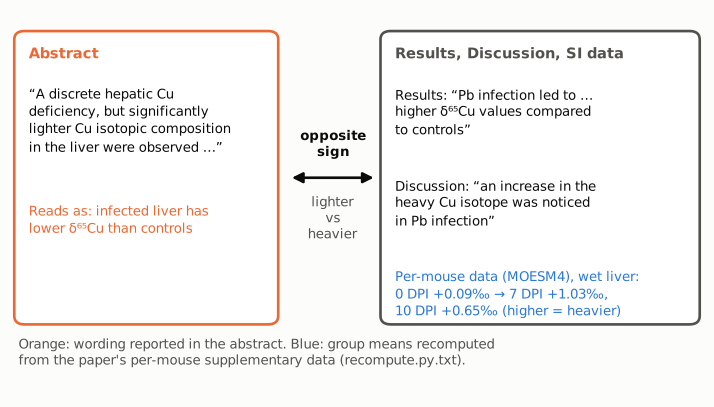
Figure 1. The conflicting statements side by side. Left (orange): the abstract sentence, which states that infected livers have isotopically lighter Cu, that is, lower δ65Cu than controls. Right: the Results and Discussion sentences on the same comparison, which state higher δ65Cu and an increase in the heavy isotope, and (blue) the wet-liver group means recomputed from the per-mouse values in the paper's Supplementary Data file. Under Eq. 3 of the paper, a higher δ65Cu means a heavier composition, so the two panels give opposite signs for one result.
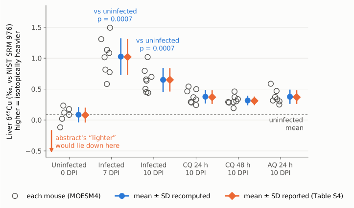
Figure 2. Liver δ65Cu (wet liver) for each mouse in the paper's Supplementary Data (open circles), with the group mean ± SD recomputed from those values (blue circles) and the mean ± SD printed in SI Table S4 (orange diamonds). The dashed line is the uninfected mean (+0.086‰). Up on this axis is isotopically heavier. Infected mice at 7 and 10 DPI all lie above all controls (exact two-sided Mann–Whitney p = 0.0007 for each). The three antimalarial-treated groups (10 DPI) lie between the untreated infected and the uninfected groups. The orange arrow marks the direction the abstract's "lighter" describes; no group of livers in the paper moves that way. The axis starts below zero only to show that direction; it does not change the visible size of any difference.
Why it matters
- A headline result has the wrong sign where most readers see it. The abstract names three hepatic isotope findings, for Fe, Cu and Zn. For Cu it states the direction opposite to the data. A reader who relies on the abstract, or on a database or review that quotes it, will record that malaria infection makes liver Cu isotopically lighter. The data show a heavier shift of about +0.9‰ at 7 DPI and +0.6‰ at 10 DPI, which is large compared with the uninfected SD of 0.12‰.
- The abstract's mechanism reads as self-contradictory. The same abstract sentence links the liver signal to "a release of Cu into the bloodstream through ceruloplasmin". The Discussion explains this release as hepatocytes exporting Cu "enriched in the light Cu isotope", which leaves the liver heavier. With "lighter" in the abstract, the stated observation and the stated mechanism point in opposite directions.
- The cross-element pattern is misdescribed. As printed, the abstract implies that liver Fe, Zn and Cu all become isotopically lighter. In the paper's data, liver Fe and Zn become lighter and liver Cu becomes heavier (Table S4), a contrast the body of the paper treats as informative about ceruloplasmin-mediated Cu export.
- What is not affected. The measurements, the group statistics, the treatment effects, the correlation of liver δ65Cu with ceruloplasmin and the conclusions in the Results, Discussion and Conclusions are all stated on the correct (heavier) basis and are internally consistent. The error is confined to one word of the abstract.
Explanations considered
- A reversed or unusual δ convention. Ruled out. Eq. 3 puts the sample's 65Cu/63Cu in the numerator, and the Methods text says explicitly that a positive δ means enrichment in the heavier isotope. The paper uses the same reading everywhere else (for example "a significant decrease in δ66Zn and δ65Cu values were observed, indicating that purified pRBCs are enriched in the lighter Zn and Cu isotopes").
- "Lighter" refers to a different compartment. Not supported. The paper does find lighter Cu in purified pRBCs (Δ65Cu = −0.88‰ against whole blood) and describes "a lighter Cu isotope signature in blood and purified RBCs". But the abstract sentence says "in the liver" and pairs the isotope finding with the "hepatic Cu deficiency", and pRBC findings are covered by the abstract's second sentence. The wet and perfused liver data contain no lighter shift.
- A different comparison, such as treated versus untreated. Not able to explain it. Treatment lowers liver δ65Cu (by 0.27 to 0.33‰ against untreated 10 DPI mice), but that is the reversal of the infection effect, and the abstract describes the infection effect ("in the liver were observed, alongside a release of Cu into the bloodstream through ceruloplasmin"). Even treated livers stay heavier than uninfected ones (+0.32 to +0.38‰ against +0.09‰).
- Wet versus perfused liver, or 7 versus 10 DPI. Not able to explain it. Both preparations and both time points shift the same way (heavier). In the perfused liver the shift is smaller (+0.35‰ with all nine infected rows, p = 0.042; +0.28‰ for the eight rows that reproduce Table S5's 0.59 ± 0.17‰). Table S5's infected mean and SD match the infected perfused group without the row the sheet labels "Liver untreated P.Berghei LR-4" (δ65Cu 1.21‰). Table S5's uninfected perfused summary (0.29 ± 0.25‰) is not reproduced by the five uninfected perfused rows either (0.31 ± 0.29‰); no subset that leaves out one row matches it, and using the population SD (0.257) would bring the SD close to the printed value but leaves the mean unexplained. The printed p = 0.0306 is not reproduced exactly by an exact two-sided Mann–Whitney test on either set of rows (0.042 with nine, 0.065 with eight). These are side points about Table S5 and does not affect the direction, which is heavier in every version.
- The aging literature cited in the Discussion. Not able to explain it. Discussion paragraph 10 mentions that "In the aging process in mice, a gradual depletion in the heavier Cu isotope in the liver is observed", citing earlier work. That sentence is about aging, not this experiment's infected-versus-control comparison.
- A text-conversion or archiving artifact. Ruled out. The conflict is in words ("lighter" against "higher δ65Cu", "increase in the heavy Cu isotope"), not in symbols or exponents, and the publisher's HTML carries the same wording as the PMC and Europe PMC copies.
- A correction already issued. None found (see Status).
Status
- Version of record. The publisher's article page (nature.com, reached through doi.org; published 3 January 2026) gives the abstract sentence "A discrete hepatic Cu deficiency, but significantly lighter Cu isotopic composition in the liver were observed, alongside a release of Cu into the bloodstream through ceruloplasmin…" and the Results sentence "…while it led to higher δ65Cu values compared to controls", word for word as in the PMC archive and the Europe PMC full-text XML. The Discussion sentences quoted above are also identical in the publisher's HTML. Every quotation was checked verbatim against the PMC-archived text with
src/show.py. - Supplements. The article lists five supplementary files. I read the Supplementary Information PDF (MOESM2: Tables S4 and S5 give liver δ65Cu means, all higher in infected mice), the Supplementary Data workbook (MOESM4: sheet "Liver" gives the per-mouse values used here, which reproduce Table S4's δ65Cu column to within 0.01‰) and the Transparent Peer Review file (MOESM1), which contains no discussion of the direction of the liver Cu shift in the abstract. The supplements resolve the question in favour of the body text: liver Cu is heavier in infection.
- Corrections and comments. As of 2026-10-24 I found no correction, erratum, retraction or expression of concern.
src/known.pyreturns no Crossref update relations, no PMC notices and no Europe PMC notices; the Crossref record has empty update-to and updated-by fields, and the publisher's page shows no Author Correction notice. The PubPeer API returned no record (HTTP 404) for the DOI, and a web search for the title together with "PubPeer" found no thread for this article. - Sources. PMC article PMC12881638; Europe PMC full-text XML; the publisher's article page at https://doi.org/10.1038/s42003-025-09464-z and its supplementary files MOESM1, MOESM2 and MOESM4; Crossref; Europe PMC; PubPeer.
Suggested correction
In the abstract, replace
A discrete hepatic Cu deficiency, but significantly lighter Cu isotopic composition in the liver were observed
with
A discrete hepatic Cu deficiency, but significantly heavier Cu isotopic composition in the liver were observed
This matches the Results (Fig. 3G: higher δ65Cu in infected wet livers), the Discussion ("an increase in the heavy Cu isotope"; "a depletion in the lighter isotope"), SI Table S4 and the per-mouse Supplementary Data. No other change to the text, figures or conclusions is needed for this point. The authors may separately wish to check the perfused-liver δ65Cu summary and p-value in SI Table S5 against the uninfected and infected perfused rows of the Supplementary Data, as noted under Explanations considered.